This article discusses the advantages and disadvantages of the various treatment options available for aortic stenosis, which is the narrowing of the valves in the heart. It will be of interest to anyone who is suffering from this form of heart disease.
Contents
- Introduction and background
- Diagnosis of aortic stenosis
- Treatment options for Aortic Valve Replacement (AVR)
- Surgical Aortic Valve Replacement
- Transcatheter Aortic Valve Implantation (TAVI)
- Conclusion
Introduction and background
The healthy heart has a number of valves which prevent the blood from flowing backwards. Normally these delicate structures are made of thin flexible and durable tissue that works efficiently over the 2000 million heart beats that occur during the course of our lives. When you consider this statistic it is not surprising that on occasions the heart valves can begin to fail. This can occur in one of two ways. Firstly, and most commonly, the valve tissue can become narrowed or stenosed. Alternatively the valves can begin to leak. This is known as regurgitation or incompetence.
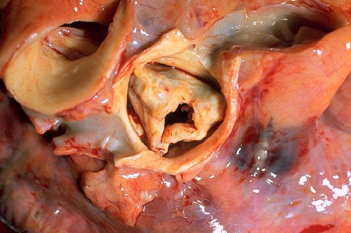
Narrowing of the aortic valve, which is known as aortic stenosis, is one of the most common forms of heart disease. It is caused by thickening, fusion and even calcification of the valve leaflets and is common in the elderly affecting over 4% of people over 80 years of age. It affects both men and woman equally. Aortic stenosis can be tolerated quite well with few symptoms for many years. However, eventually symptoms of breathlessness on walking, angina or collapse may develop. When this happens it is important to get treatment quickly as untreated symptomatic aortic stenosis is progressive and often fatal.
Diagnosis of Aortic Stenosis
Aortic stenosis typically causes a loud heart murmur that your doctor can hear using a stethoscope on your chest. This heart murmur is produced by the turbulent flow of blood being squeezed through the narrow heart valve. The diagnosis is confirmed by a heart ultrasound or echocardiogram. This test uses sound waves to visualise the heart muscle and valve and determine the severity of the narrowing. Other tests that might be needed include an ECG and coronary angiogram to assess whether there is any narrowing in your coronary arteries as well. Once you have had these tests your cardiologist can advise you on the most appropriate treatment for you.
Treatment Options for Aortic Stenosis and TAVI
Unfortunately aortic stenosis cannot be treated with medicines and in the majority of cases it is better to relieve the narrowing of the valve. Traditionally this has been exclusively by open heart surgery to remove the narrowed valve and replace it with a new man-made valve. Recently, however, a new minimally invasive technique has been developed that allows a new valve to be implanted in the heart through a tube, known as a catheter, inserted through a blood vessel. This treatment is known as Transcatheter Aortic Valve Implantation or TAVI for short.
TAVI has the advantage of avoiding the need to open the chest and the use of a heart-lung bypass machine, and therefore has a quicker recovery and return to normal activities time. The recommendation of which treatment is best for you is usually determined by a ‘multidisciplinary’ heart team which may include many doctors including cardiologists, cardiac surgeons and anaesthetists. The heart team will consider, amongst other things, the severity of your symptoms and aortic stenosis and other illnesses you may have that would increase the risk or reduce the benefit of one or other treatments.
Occasionally, the narrowed heart valve can be simply stretched using a balloon (balloon aortic valvoplasty or BAV) or no intervention may be recommended. Below we describe, in more detail, the various options for treating aortic stenosis and outline the pros and cons of each treatment option.
Surgical Aortic Valve Replacement (AVR)
Aortic valve replacement by open heart surgery is the standard way of replacing a diseased aortic valve. This involves opening the chest wall through the breast bone to get access to the heart. The heart is stopped using a special solution which protects the heart and the diseased valve removed and a new man-made valve sewn in its place. While the heart is stopped the circulation is supported by a heart-lung bypass machine.
There are two main categories of valves, namely, biological and mechanical. The optimal choice is dependent on many factors including age, life style and the presence of other medical conditions. Biological valves are usually made from pig valve tissue (porcine) or cow pericardial tissue (bovine). These valves are easy to implant, durable (lasting from 15 to 20 years), and may allow patients to avoid lifetime use of anticoagulants (blood thinning medications). However, in younger patients (aged below 60), these valves are likely to wear out and will need replacing at some stage.
Mechanical valves are very durable and may last for many years but do require the patient to take anticoagulants for life. After surgery most patients will stay in hospital for one week and then continue their recovery over a 2-3 month period.
Advantages of Surgical Aortic Valve Replacement (AVR)
Surgery is very effective at replacing the diseased aortic valve with excellent results. There are many different types of valve than can be chosen to suit the individual patient. In many cases, patients may have other heart conditions like coronary disease or disease of other heart valves and surgical aortic valve replacement can be combined with coronary artery bypass grafting or other valve surgery if necessary.
Disadvantages of Surgical Aortic Valve Replacement (AVR)
In healthy patients isolated surgical AVR is a relatively low risk operation. Other factors such as the patient’s age and presence of lung or kidney disease can considerably increase the risk and length of recovery time after surgical AVR. Repeat heart surgery or combined surgical AVR and other operations can also increase the risk. If the risk is high then other forms of treatment may be considered (see below). The recommended treatment option should be based on a multidisciplinary team (cardiac surgeon, cardiologist, anaesthetist and others) decision taking in to account all factors including patient's preference.
Non-Surgical Aortic Valve Replacement -
Transcatheter Aortic Valve Implantation (TAVI)
In TAVI a new aortic valve is implanted in your heart via a tube or catheter. This tube is normally introduced through the artery at the top of your leg (transfemoral approach) and then advanced up to the heart through the blood vessels. If these blood vessels are narrowed or diseased then the catheter can be introduced via the blood vessel under your collar bone (subclavian approach) or directly into the aorta by making a small incision in your chest wall (direct aortic approach). The final option is to introduce the catheter directly into the heart from the left side of the chest. This is known as a transapical approach.
For further information on TAVI and other non-surgical treatments for Aortic Stenosis - see Transcatheter Aortic Valve Replacement - Latest Treatments for Aortic Stenosis
Conclusion
Aortic stenosis is the most common form of valvular heart disease and if untreated is often fatal. Timely intervention to replace the valve can both improve symptoms and increase lifespan and therefore, in general, should not be delayed once symptoms develop. There are many treatment options for patients including open heart surgery and transcatheter aortic valve implantation (TAVI). A multidisciplinary heart team is essential to consider the most appropriate treatment for each patient.