New imaging techniques for the assessment of heart disease in patients with diabetes


This article emphasises how important it is to accurately diagnose heart problems in diabetic patients. Two experts in cardiovascular disease and diabetes have come together to advise on what the best methods are to spot potential heart disease in diabetic patients.


Contents

Overview of diabetes and Coronary Artery Disease (CAD)

Diabetes is reaching epidemic proportions in both the Western and developing worlds. In 2009 the Quality of Outcomes Framework wrote that there were 2.6 million people in the UK reported to have diabetes. This figure is set to reach 4 million by 2025. Due to the increasing prevalence of obesity, the incidence and vascular complications of type 2 diabetes will continue to rise (The Foresight Report estimates that by 2015, 64% of adults aged 21 to 60 in the UK will be obese, and by 2025 this will rise to 83%).

Nearly 80% of patients with type 2 diabetes suffer from cardiovascular illness, with coronary artery disease (CAD) representing the major component of this disease. Approximately 10–15% of patients admitted to hospital with a heart attack (myocardial infarction) and 20% of patients dying from coronary heart disease (CHD) have diabetes as a contributing factor. In general, diabetic patients have more extensive atherosclerosis with a higher prevalence of multi-vessel CAD and frequent silent heart attacks (without chest pain). Some studies have even suggested that diabetic patients without CAD have the same risk for future cardiac death as non-diabetic patients with established CAD1 (1). Cardiovascular mortality and morbidity appear significantly worse in those with diabetes than those without. Diabetes is therefore placing a large burden on our health care system, both directly and indirectly.

Screening for Coronary Artery Disease

There are a number of screening methods for the diagnosis of coronary artery disease and a number of limitations with the current screening methods. The main current screens are as follows:

ECG / Exercise ECG

Electrocardiography (ECG) is the first test, and it records the electrical activity of the heart. It is painless and harmless and takes only a few minutes. The exercise ECG entails the patient on a treadmill or an exercise bicycle with leads being attached to the chest that measure how the heart functions whilst exercising. Abnormal ECGs / Exercise ECGs in diabetic patients are associated with a high risk of heart attacks. However, diabetic patients may have peripheral neuropathy which may limit exercise, or result in a poor heart rate response to exercise due to autonomic dysfunction. Thus these tests have poor sensitivity and specificity which make them a poor screening tool. The use of an exercise test alone has led to many false positive tests leading to unnecessary, expensive and invasive coronary angiography tests, in which nearly 50% of the resulting tests were normal. However, the exercise ECG test remains a very useful investigation in good hands.

Coronary Artery Calcium Scoring

All the office-based screening calculations assess a patient’s 10-year risk of developing coronary heart disease. Over the last few years, coronary artery calcium (CAC) imaging, where a cardiac CT scan reveals information about the extent of hardened plaque in the coronary arteries (atherosclerosis), has been increasingly used to further assess a patient’s risk, particularly those in an intermediate risk category. However, there is a poor correlation between CAC and the degree of narrowing of the coronary blood vessels and its ability to predict which patients will go on to suffer a medical event, although there has been a large amount of data demonstrating its utility in predicting cardiovascular survival over 7 – 10 years. Patients who are without symptoms and assigned to the high-risk group with office-based risk assessment calculations will benefit from intensive risk modification. In asymptomatic diabetic subjects Raggi and colleagues  (2) have shown that mortality rates are significantly higher than their non-diabetic counterparts with similar CAC scores. The possible explanation is probably due to a faster progression of atherosclerosis.
 
Patients in the low risk group are advised to follow a healthy lifestyle and their risk factors should be treated as and when diagnosed according to current guidelines. Those in the intermediate-risk group pose a clinical dilemma and it is this group of patients who will benefit most from CAC scoring. The management strategy for patients who are classified in the intermediate risk category remains predominantly subjective, therefore using the CAC score gives us a tool to try to re-classify some of these patients into higher or lower risk groups making management decisions easier and more evidence-based.

The above graph shows that for every increase in calcium score there was a greater increase in mortality for diabetic compared to non diabetic subjects. Also for the same calcium score diabetic patients are at a higher risk of death than non-diabetic.

CTCA (CT Coronary Angiography) Technique

CT Coronary Angiography (CTCA) requires the injection of an iodine-based substance into the patient through a large vein and then x-ray type images are taken. The actual scan time is less than 15 seconds in over 90% of patients. The image quality can be improved with the use of a special spray that is administered under the tongue approximately five minutes prior to the scan and also beta-blockers can be administered to get an ideal heart rate of less than 65 beats per minute.

Clinical role of CTCA

Non-invasive coronary angiography by computed tomography (CTCA) is a new and evolving technique which can accurately image/detect the presence of coronary artery disease (atherosclerosis) inside the heart arteries. Unlike the two-dimensional image of conventional invasive angiography, this technique also allows us to visualise the vessel wall and the plaque itself, and has the potential for plaque characterisation, bringing us a step closer to identifying the characteristics of a vulnerable plaque (plaque prone to rupture), which is the ‘holy grail’ of cardiology. It is able to pick up "silent" disease, which is especially important in diabetic patients.

In order to get high quality diagnostic images of the heart in a reproducible manner, newer scanners such as the Dual-source CT scanner are better suited for imaging the coronary arteries. Through a combination of Dual-source CT-coronary angiography (DS-CTCA) and the measurement of specific biomarkers, which are biochemical substances produced by the body that allow us to measure the progress of a disease, we are able to assess not only the total coronary artery disease plaque burden, but also the ‘stability’ of the plaque and can possibly identify those people who are at a higher risk of rapid progression of the disease.

Myocardial Perfusion Scan (MPS)

The Myocardial Perfusion Scan (MPS) is a well-established test which provides important prognostic information to stratify cardiac patients according to their risk. It is a scanning procedure that is able to illustrate the function of the heart muscle (myocardium). There is an extensive body of evidence that has accumulated over the last 2–3 decades regarding the usefulness of MPS, which allows us to see how well blood is reaching the heart muscle through the coronary arteries, in the diagnosis and short term prognosis of coronary artery disease. The limitation of this technique arises from the fact that it is necessary for there to be significant (>50%) narrowing of blood vessels causing relative discrepancies in blood flow to the heart.
MPS cannot, therefore, be used as a primary screening tool. A normal MPS does not provide any information regarding the extent of the plaque burden of the coronary arteries per se. Though MPS accurately predicts the severity of obstructed coronary arteries, it does not directly image the heart arteries as can be done now by new and evolving Cardiac CT technology such as the CTCA.

Despite these advances nearly 50% of patients with coronary artery disease will not receive medical attention before suffering a heart attack that may result in death. This is probably attributable to the fact that nearly two thirds of culprit plaque lesions that are responsible for heart attacks/death are not known to cause any significant coronary narrowing (>50% stenosis) and are identifiable by current CTCA techniques.

CAC and MPS (Coronary Artery Calcium score and Myocardial Perfusion Scan)

Clinical studies have shown that combining Coronary Artery Calcium Imaging and Myocardial Perfusion Scanning provides an excellent method of detecting those people at a high risk of cardiac events (through death, heart attacks etc) (3).

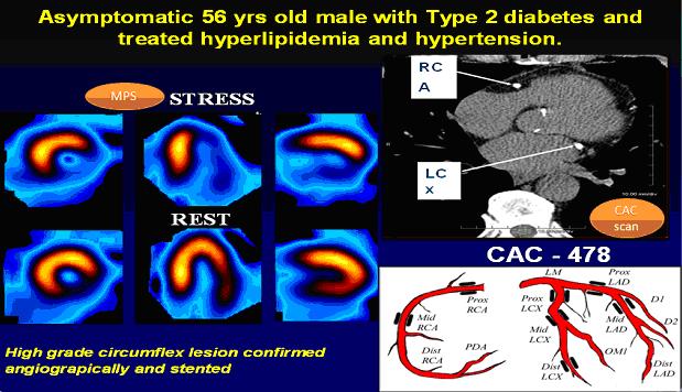
These pictures show how well new technology guides us in making the best decisions on the management of patients. A 56 year old gentleman known to have diabetes, hypertension and hypercholesterolaemia was without any symptoms but underwent a Coronary Artery Calcium (CAC) scan as part of initial screening; this revealed a high calcium burden (CAC - 478). So we arranged for him to have a Myocardial Perfusion Scan (MPS) that demonstrated a significant compromise in blood flow to the heart muscle. He was then successfully treated with invasive coronary angiogram and stent insertion. Click on image to enlarge

NICE guidelines for chest pain

The NICE (National Institute for Health and Clinical Excellence) Guidelines relating to “Chest Pain” (April 2010) have provided clear-cut guidelines regarding the use of cardiac CT (both calcium imaging and CT coronary angiography) in combination with Myocardial Perfusion Scanning when required. We recently concluded a large clinical trial (RADICAL) to assess the clinical and cost-effectiveness of Dual-source CT coronary angiography in patients from three NHS hospitals referred to the chest pain clinics. These patients are recruited at the NHS site and then sent to the Wellington Hospital for CTCA and MPS (if needed). The DS-CTCA is combined with Myocardial Perfusion scanning when intermediate narrowing of the coronary arteries is noted on the CTCA to guide management strategies.
 
Interim results are very encouraging and suggest that:
  • 2/3 patients who had CT angiogram at the Wellington did not require further testing
  • Most patients preferred CT angiogram compared to invasive cardiac angiogram
  • Initial results from CT angiogram proved useful in deciding method of treatment

Conclusion

In summary, diabetic patients have a four times greater chance of developing silent coronary artery disease compared to the general population. By 2020 there will be over 400 million diabetics worldwide, 70-80% of whom will suffer cardiovascular problems and coronary artery disease is estimated to become the number one killer worldwide (according to the World Health Organization). It is therefore imperative that methods of early detection of coronary artery disease are established to combat this growing threat.

References:

1. Haffner SM, Lehto S, Rönnemaa T, Pyörälä K, Laakso M. Mortality from coronary heart disease in subjects with type 2 diabetes and in nondiabetic
subjects with and without prior myocardial infarction. New England Journal of Medicine 1998;339(4):229–234.

2. Raggi P, Shaw LJ, Berman DS, Callister TQ. Prognostic value of coronary artery calcium screening in subjects with and without diabetes. Journal of the American College of Cardiology 2004;43(9):1663-1669.

3. Anand DV, Lim E, Hopkins D, Sharp P, Fourie S, Raval U, Lipkin D, Corder R, Lahiri A. Prevalence and clinical predictors of silent myocardial ischaemia in patients with type 2 diabetes. Heart 2004;90(Supp. II):A58.
An X-ray image of the blood vessels following the injection of a dye to improve visibility. Full medical glossary
X-ray imaging of the blood vessels following the injection of a dye to improve visibility. Full medical glossary
A blood vessel that carries blood away from the heart. Apart from the pulmonary artery and umbilical artery, all arteries carry oxygenated blood. Full medical glossary
Disease leading to fatty deposits in the inner walls of the arteries, which reduce and may eventually obstruct blood flow. Full medical glossary
The part of the nervous system supplying muscles such as the heart and bowels, known as involuntary muscles. Full medical glossary
One of a group of drugs that block beta-receptors to slow the heart rate, or constrict the airways and arteries. Full medical glossary
A group of drugs that block beta-receptors to slow the heart rate, or constrict the airways and arteries. Full medical glossary
A substance that can be measured to help healthcare professionals to assess normal processes, disease processes or a person's response to treatment. Full medical glossary
A fluid that transports oxygen and other substances through the body, made up of blood cells suspended in a liquid. Full medical glossary
An element that forms the structure of bones and teeth and is essential to many of the body's functions. Full medical glossary
Relating to the heart Full medical glossary
Disease of the heart and blood vessels, usually due to atherosclerosis. Full medical glossary
The basic unit of all living organisms. Full medical glossary
A substance present in many tissues and an important constituent of cell membranes although high concentrations of a certain type of cholesterol in the blood are unhealthy. Full medical glossary
A condition that is linked to, or is a consequence of, another disease or procedure. Full medical glossary
Relating to the arteries supplying the heart itself. Full medical glossary
A technique for X-ray imaging of the coronary arteries, which involves introducing a catheter through a blood vessel and threading it towards the heart, then injecting a contrast medium to improve visibility. Full medical glossary
Narrowing of the blood vessels supplying the heart muscle, leading to symptoms such as angina and sometimes to a heart attack or myocardial infarction Full medical glossary
The abbreviation for computed tomography, a scan that generates a series of cross-sectional x-ray images Full medical glossary
A disorder caused by insufficient or absent production of the hormone insulin by the pancreas, or because the tissues are resistant to the effects. Full medical glossary
The process of determining which condition a patient may have. Full medical glossary
An abbreviation for disseminated intravascular coagulation. Full medical glossary
The abbreviation for electrocardiogram, a tracing of the electrical activity of the heart to help in the diagnosis of heart disease. Full medical glossary
A sudden outbreak of infection that affects a large proportion of a population. Full medical glossary
The basic unit of genetic material carried on chromosomes. Full medical glossary
An organ with the ability to make and secrete certain fluids. Full medical glossary
The death of a section of heart muscle caused by an interruption in its blood supply. Also called a myocardial infarction. Full medical glossary
High levels of cholesterol in the blood. Full medical glossary
High blood pressure. Full medical glossary
intermittent claudication Full medical glossary
The number of new episodes of a condition arising in a certain group of people over a specified period of time. Full medical glossary
Death of a piece of tissue owing to obstruction of its blood supply. Full medical glossary
Death of a portion of tissue due to inadequate blood supply Full medical glossary
Insufficient oxygenation to a part of the body due to poor blood supply. Full medical glossary
a general term to cover any abnormality such as a wound, infection, abscess or tumour. Full medical glossary
Tissue made up of cells that can contract to bring about movement. Full medical glossary
Death of an area of heart muscle due to poor blood supply. This is commonly known as a heart attack. Full medical glossary
Damage to or disease of nerves. Full medical glossary
Excess accumulation of fat in the body. Full medical glossary
Any flat, raised patch; for example, a raised patch on the skin, fatty deposit in the inner wall of an artery, or layer over the surface of a tooth. Full medical glossary
An assessment of the likely progress of a condition. Full medical glossary
Lying face-downwards. Full medical glossary
rheumatoid arthritis Full medical glossary
A way to identify people who may have a certain condition, among a group of people who may or may not seem to Full medical glossary
Narrowing of a tubular structure or valve. Full medical glossary
A tube placed inside a tubular structure in the body, to keep it patent, that is, open. Full medical glossary
An abbreviation for tricyclic antidepressant drug. Full medical glossary
Relating to blood vessels. Full medical glossary
A blood vessel that carries blood towards the heart. Full medical glossary
Relating to the sense of sight (vision). Full medical glossary